Detailed · Deep study
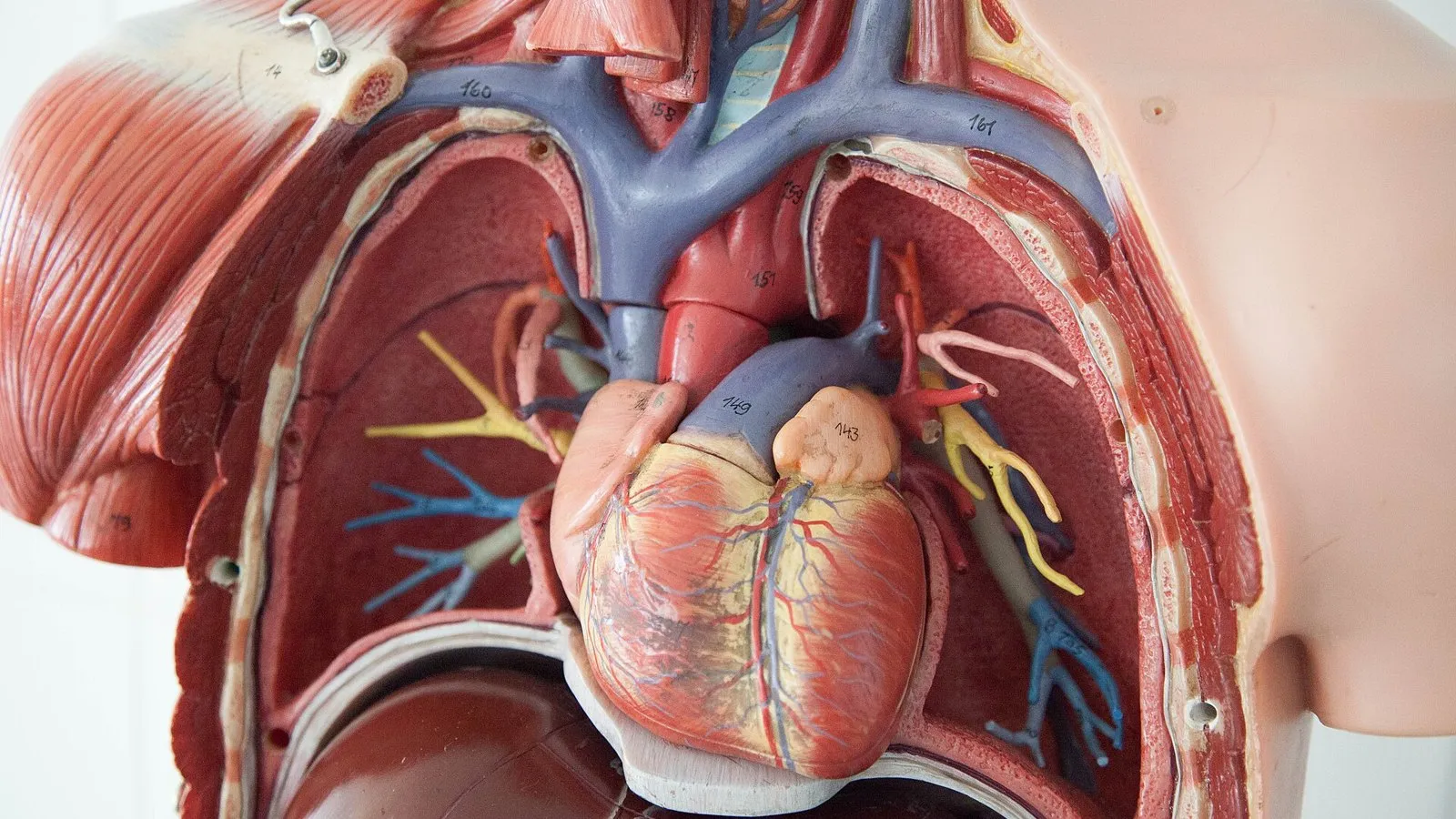
1Structure of the mammalian heart
| Structure | Description and role |
|---|---|
| Right atrium | Receives deoxygenated blood from the body through the vena cava |
| Right ventricle | Pumps deoxygenated blood to the lungs through the pulmonary artery |
| Left atrium | Receives oxygenated blood from the lungs through the pulmonary vein |
| Left ventricle | Pumps oxygenated blood to the body through the aorta |
| Septum | Muscular wall dividing the left and right sides |
| Muscular wall | Made of cardiac muscle, which contracts to pump blood |
| One-way valves | Open only one way, so blood cannot flow backwards |
| Coronary arteries | Branch from the aorta over the surface of the heart, supplying the heart muscle with oxygen and glucose |
Blood is pumped away from the heart in arteries and returns to the heart in veins.
2Wall thickness and the septum (Supplement)
| Comparison | Explanation |
|---|---|
| Left ventricle wall is thicker than the right ventricle wall | The left ventricle pumps blood at high pressure all the way round the body. The right ventricle pumps blood only to the lungs, which are close by; a lower pressure there also avoids damaging the delicate lung capillaries |
| Atria walls are thinner than ventricle walls | The atria only push blood a short distance, into the ventricles just below them, so they need less muscle |
The septum keeps oxygenated blood (left side) separate from deoxygenated blood (right side). If they mixed, blood going to the body would carry less oxygen, so less oxygen would reach respiring cells.
3How the heart pumps (Supplement)
- Blood flows into both atria from the veins (vena cava and pulmonary vein).
- The atria contract. Blood is pushed through the open atrioventricular valves into the ventricles.
- The ventricles contract. Pressure rises and the atrioventricular valves shut, preventing backflow into the atria.
- The semilunar valves open and blood is forced into the pulmonary artery and aorta.
- The ventricles relax. The semilunar valves shut, preventing backflow from the arteries into the ventricles.
4Monitoring the heart and exercise
| Method | What it shows |
|---|---|
| ECG (electrocardiogram) | Electrical activity of the heart, from electrodes on the skin; a trace on a screen |
| Pulse rate | Beats per minute, felt where an artery passes near the skin, e.g. the wrist or neck |
| Listening to the valves with a stethoscope | The sounds of the valves closing; abnormal sounds can show a faulty valve |
Practical
Effect of physical activity on heart rate
Aim. To investigate how physical activity affects heart rate.
Method
- Sit quietly for 5 minutes, then count your pulse for 15 seconds and multiply by 4 (beats per minute). Repeat to find a mean resting rate.
- Do a set activity, e.g. step-ups for 2 minutes.
- Immediately measure your pulse rate again.
- Measure it every minute afterwards until it returns to the resting rate (recovery time).
- Repeat with a more intense or longer activity.
| independent variable | Intensity or duration of physical activity |
|---|---|
| dependent variable | Heart rate (beats per minute) |
| controlled variable | Same person, same type of activity, same method of counting, same position when measuring |
Results. Heart rate increases during exercise, and more intense exercise gives a higher rate. After exercise it falls gradually back to the resting rate.
Safety. Do not take part if you have a medical condition that makes exercise unsafe. Stop if you feel unwell.
5Coronary heart disease
In coronary heart disease (CHD), the coronary arteries become blocked. Fatty deposits build up in the artery wall and narrow the lumen; a blood clot can then form and block it completely. The heart muscle beyond the blockage receives no oxygen or glucose, so it cannot respire and may die. This is a heart attack.
| Risk factor | Why it increases risk |
|---|---|
| Diet | A diet high in saturated fat and salt raises blood cholesterol and blood pressure |
| Lack of exercise | Increases the chance of obesity and high blood pressure |
| Stress | Can raise blood pressure |
| Smoking | Chemicals in smoke damage artery walls and make clots more likely |
| Genetic predisposition | Some people inherit a higher risk |
| Age | Risk increases with age |
| Sex | Males are at greater risk than females of the same age |
| Reducing the risk | How it helps |
|---|---|
| Diet: eat less saturated fat, salt and sugar; more fruit, vegetables and fibre | Lowers blood cholesterol, blood pressure and the chance of obesity, so fewer fatty deposits form |
| Exercise: regular physical activity | Strengthens the heart, lowers blood pressure, helps control body mass and improves the balance of blood fats |
6Exam focus
Practice questions
Quick self-check
Answer in your head first, then reveal.
Supply the heart muscle with (oxygenated) blood / oxygen and glucose.
Aorta.
Heart rate increases during activity; it decreases gradually back to the resting rate afterwards.
Any four of: diet high in saturated fat, lack of exercise, stress, smoking, genetic predisposition, age, sex.
The left ventricle pumps blood all round the body / further; it needs a higher pressure (the right ventricle pumps only to the lungs).
Atrioventricular valves close; preventing backflow into the atria; semilunar valves open so blood flows into the arteries.
Muscles respire faster / need more energy; need more oxygen and glucose (and remove more carbon dioxide); a faster heart rate delivers blood more quickly.